We use cookies to ensure that we give you the best experience on our website.
Image processing and data analysis
Get the most out of your imaging data
Image processing and data visualisation service
Need help with your existing data? At X-Ray Labs, we perform three-dimensional tomographic reconstruction of the data generated from third-party micro-CT systems, synchrotron tomography (including phase retrieval in phase-contrast imaging and tomography), neutron tomography, and optical projection tomography. We cover both parallel-beam and cone-beam geometry, as well as other arrangements such a s laminography, using conventional and algebraic reconstruction techniques. We also develop custom image processing scripts for both 2D and 3D datasets, for instance data normalisation, artifact correction, segmentation, deconvolution, and quantitative analysis of volume data. In addition, we generate beautiful 3D visualisations of your data using volume rendering, and we can export your tomographic dataset into a volume mesh compatible with CAD design software.
Examples of our service include:
2D & 3D segmentation
Quantitative analysis
Mesh generation & smoothing
Isosurfacing
Tomography reconstruction and artifact reduction
Image stitching, registration and data fusion
Denoising and feature extraction
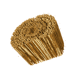
Gallery
X-ray micro-CT of a beetle. After reconstruction, we performed denoising and histogram segmentation into intensity clusters. Finally the data was rendered according to the simplified intensity levels.
X-ray micro-CT of a chocolate bar. The data was reconstructed and smoothed to aid visualisation. Surface rendering was then performed on the intensity histogram rescaled to 8-bit.
X-ray micro-CT of a marshmallow for a porosity study. The data was reconstructed and denoised in order to improve the visibility of the fine porosity. Surface rendering was then performed on the intensity histogram rescaled to 8-bit.
Neutron tomography of a porous sample. The image was reconstructed using a cone-beam tomography reconstruction algorithm. It was then segmented and the pore size distribution was extracted from the segmented data.
Quantitative analysis
Often, you might need to gather quantitative information from a micro-CT scan. For instance you may be interested in the particle size distribution of a composite sample, the pore size distribution of a concrete sample, the wall thickness of a metal component, and so on. This type of quantitative information can be extracted from micro-CT calibrated data. This process however can be extremely tedious or outright unfeasible for large datasets. This is where scripting and automation can be used. At X-Ray Labs, we have expertise in developing scripts and automated routines for quantitative data extraction from micro-CT and microscopy datasets.
Examples of our expertise include:
Defect analysis
Topology optimisation
Material segmentation
Composite analysis
Morphological analysis in 2D & 3D
Network analysis of porous or interconnected samples
Gallery
Micro-CT scan of a wooden stem with visible vascular bundles (the dark regions in the top-left image). Image acquired with 10 micron pixel size. Total scan duration: 30 minutes.
The vascular bundles can be segmented based on the histogram and their size extracted from the segmented data. Specifically, the cross-sectional area of each bundle can be measured. Assuming a circular cross-section, the radius can then be quantified.
Either the original reconstructed volume, or the segmented volume can be rendered using intensity information. Volume rendering does not provide quantitative information, but it is extremely useful to glance at the main structure of the sample.
Third-party data analysis
One of the key issues with modern industrial CT and micro-CT, is the presence of image artifacts. Artifacts are artificial structures appearing in the digital reconstruction of the sample data, which do not corresponds to real structures in the sample. Artifacts arise from a variate of causes, for instance sample misalignment, noisy data (the main culprit for ring artifacts), polychromatic beam (so called “beam hardening” artifacts), or highly-absorbing structures in the sample (similar to “metal” artifacts well known to medical CT practitioners).
Artifact mitigation or removal is one of the service that we provide to users who have already acquired micro-CT data, for instance from a synchrotron facility. Artifact correction can be approached from the original data or from the reconstructed data, with a variety of techniques. Artifact reduction is often the critical step to be able to extract quantitative information from the sample scan.
We develop the entire data-processing workflow for third-party data: pre-processing, reconstruction, segmentation, analysis and visualisation. In addition to standard image processing routines, we have deep expertise in X-ray phase-retrieval algorithms useful with synchrotron phase-contrast X-ray imaging data. Phase retrieval enables contrast enhancement and noise reduction in low-absorbing samples, thus mitigating the presence of artifacts in this type of scans.
Examples of our service for third-party data include:
Noise reduction (denoising)
Image stitching (for partial scans)
Beam-hardening artifacts corrections
Ring artifacts correction
Partial volume effect
Scan misalignment correction
Phase retrieval algorithms
Image segmentation and analysis
Gallery
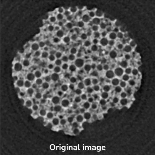
Micro-CT reconstruction of a concrete sample. Ring artifacts and beam-hardening artifacts are visible in the raw reconstruction.
Corresponding image with ring artifacts removed. The beam-hardening is however still present, producing a degree of blurring in the image.
After beam-hardening correction, the image is as crisp as the resolution of the system allows, allowing for unbiased quantitative parameter estimation.
Get in touch
Do you have any other questions on our image processing and data analysis services?
Fill this brief contact form and we will be in touch within 1 business day.